


National event highlights Norwich’s leading role in 4D heart imaging
Our cardiology and radiology team have highlighted Norwich’s role as a leading place for state-of-the-art cardiac imaging at a national webinar.
Patients at the Norfolk and Norwich University Hospital were the first in the country to benefit from detailed 4D flow images of the heart using specialist magnetic resonance imaging (MRI). Since 2021, around 1,500 patients have received 4D heart imaging, which is helping to improve clinical understanding of pressure changes in the heart and to help diagnose heart valve disease earlier.
The 4D Lab team led a national webinar run by the British Society of Cardiovascular Magnetic Resonance, which was attended by more than 120 clinicians from across the country to learn about the use of this imaging technique.
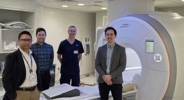
The presentation was led by MRI Radiographer Kurian Thampi, Dr Pankaj Garg, Honorary Consultant Cardiologist and Clinical Scientist in Cardiovascular Medicine at the University of East Anglia (UEA) and Cardiology Specialty Registrar Dr Alexander Gall to highlight how 4D flow imaging has moved most quickly from research towards clinical practice to diagnose valvular heart disease. This webinar covered 4D flow measures, what is required to acquire a reliable dataset and how the resulting measurements can be used in the assessment of patients with valve disease.
Dr Garg said the webinar has led to the team advising other hospitals in the country after successfully establishing 4D imaging at the NNUH.
“We are leading the way Norwich with specialists in 4D imaging at NNUH, which is being recognised locally, regionally and nationally, as well as dedicated cardiac imaging at the Community Diagnostic Centre (CDC) and we have radiographers trained in advanced cardiac imaging techniques, including 4D Flow MRI.”
“We have cut the average length of a scan down to between eight and ten minutes and we now have access to huge data sets and using 4D flow data for around 1,500 patients. Ultrasounds didn’t give us a map of pressures in the heart and we have not been able to see this level of imaging and the pressure flows in the heart before.”
The innovative work has been made possible thanks to the purchase of a dedicated cardiac MRI machine at the CDC, which was funded following fundraising by the Norfolk Heart Trust (NHT) and support from the N&N Hospitals Charity.
Since starting the work in Norwich five years ago, Dr Garg has been joined by Dr Ciaran Clarke, Clinical Research Fellow at UEA and junior cardiology registrar, funded by the British Heart Foundation focusing on using 4D flow imaging to analyse hypertension flow in the right heart.
Dr Gall, who is also an Advanced Cardiac Imaging Research Fellow at UEA, is funded by Edward Lifesciences and is focusing on the right heart and right arterial flow.
Rebekah Girling, Clinical Services Research Lead and NIHR Imaging Specialty Lead for East of England, added: “This is an excellent example of how clinical expertise, research and innovation can come together to advance patient care. Kurian’s Diagnostic Radiographer contribution to a national webinar demonstrates the depth of specialist expertise being developed at NNUH, while the 4D Flow MRI work led by Dr and the 4D Lab team shows how imaging research can translate into new approaches to understanding heart disease. I am particularly proud to see our teams not only delivering innovative research locally, but also sharing their expertise nationally and helping shape the future of imaging research and clinical practice.”