Radiotherapy Physics and Nuclear Medicine
If you’re interested in physics, these are the disciplines of Healthcare Science that you should follow.
Watch the videos below to learn more about the specialty, including the treatment of cancer patients using ionisation radiation and what they do in their daily role.
Alison Vinall, Consultant Clinical Scientist and Head of Radiotherapy Physics, explains the role of a Radiotherapy Physics Scientist:
Will Holmes-Smith, Lead Clinical Scientist, talks about QA and Machine Dosimetry:
Vicki Currie, Lead Clinical Scientist, explains treatment planning and Brachytherapy:
Anthi Alexandrou, Clinical Scientist, explains treatment planning:
Susannah leah, Trainee Clinical Scientist, talks about the training route:
Nuclear Medicine is the science of using radioactive substances introduced into the body for diagnosis and treatment of disease.
Healthcare Scientists in nuclear medicine work alongside doctors and nurses to undertake functional imaging (seeing how parts of the body are working) by using a radiation source introduced into the body through the application of radiopharmaceutical (a radioactive chemical).
Treatment of cancers through application of radiopharmaceuticals to destroy the cancer cells is also a major part of their work and clinical scientists ensure all procedures are performed safely and with minimum radiation exposure by calculating doses required.
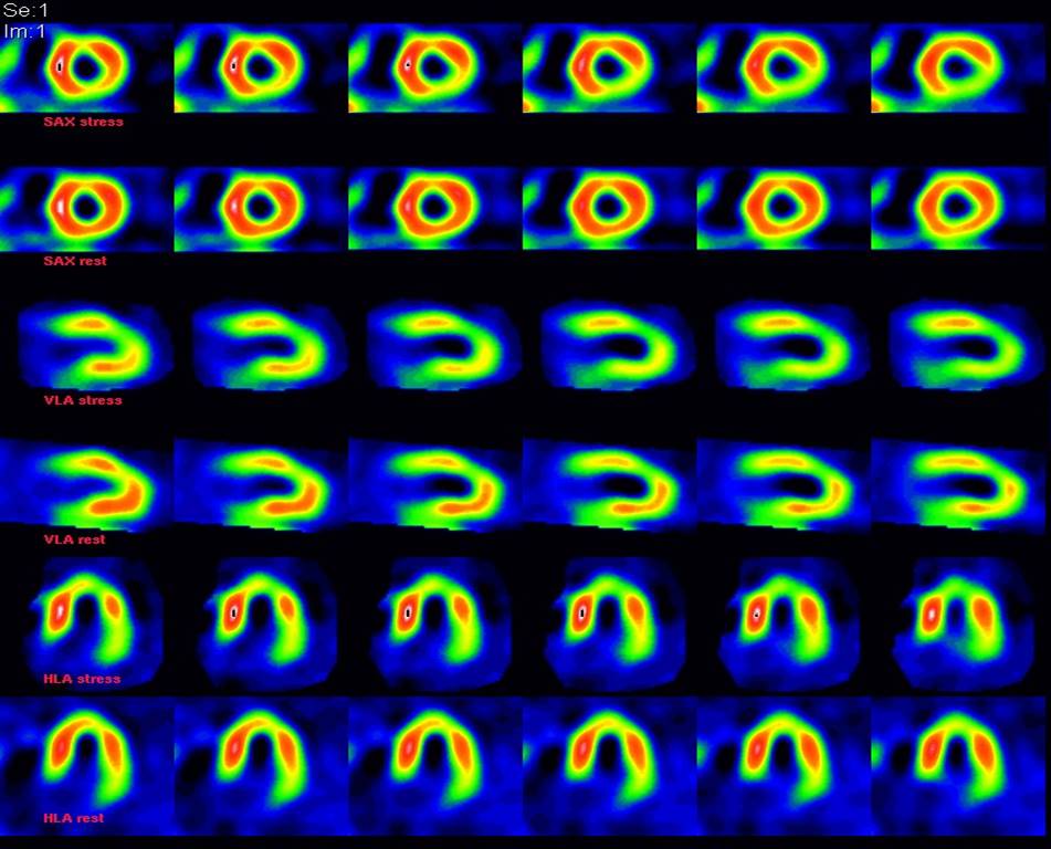
These images show blood flow around the heart to identify areas of healthy/unhealthy heart tissue to help diagnose heart disease.

These images show the difference in blood flow within the brain for a patient with depression and after recovery.

In 2019, scans included:
- 5,771 administrations in total
- 37 different procedures
- 2,200 bone scans
- 830 sentinel node
- 520 kidney scans
- 1,100 heart scans
- 200 thyroid scans
- 223 parathyroid scans
- 184 tracer studies
- 150 scans for cancer detection.

We have a PET/CT scanner which detects the radioactivity emitted from the body after the radiopharmaceutical has been injected. It takes images in slices and then creates a 3D image. PET scanner image
PET scans are used to investigate lung carcinoma, colorectal cancer, lymphoma, melanoma, neuroendocrine/pancreatic cancer, the efficacy of anti-cancer drugs, healthy/unhealthy heart tissue in patients with known severe heart disease or infection and brain function.
By putting together the functional image (PET) and the anatomical image (CT), we create a hybrid image which can help the doctor identify the presence and site of cancer.
We have three new Gamma Cameras with Multi-Slice Diagnostic Quality CT which gives much better images.
An important role of the clinical scientist is to introduce new technology that helps improve imaging and treatment while reducing exposure to radiation.


